Written by: Prof. Dr. Semih Halezeroğlu / Head of the Department of Thoracic Surgery, Acıbadem University Faculty of Medicine
In this article, detailed information is provided about what **Pleural Mesothelioma** is, how it occurs, its symptoms, and what international sources recommend. Based on this information, you can find detailed information about the diagnosis, treatment, surgical methods, surgical risks, and postoperative care for mesothelioma.
Pleural Mesothelioma
Mesothelioma is a malignant tumor that arises in the pleura (lung lining). Our lungs, diaphragm, the inner surface of the ribs, and the mediastinum are covered by a membrane. This membrane is called the pleura. This very thin membrane prevents these organs and structures from sticking together, separating them. Occasionally, non-benign tumors can arise in this pleura, which is known as pleural mesothelioma in medical terminology. There is also a type of mesothelioma that arises in the peritoneum, which covers the inside of the abdomen. In this article, we will only discuss pleural mesothelioma.
How Does Mesothelioma Occur?
It has been established for many years what causes mesothelioma, or the factors that lead to its development. Previously, there was a substance used in industry (such as in water pipes, ports, the maritime industry, large buildings, and constructions) called ASBESTOS. This substance was the primary factor that led to the emergence of mesothelioma. Although asbestos use is banned in the industry today, the disease has not been completely eradicated. Why? Because the substance known as asbestos shares similar characteristics with a mineral found in some volcanic areas in the soil, which is called ERIONITE.
In fact, in our country, especially in the Central and Southeastern Anatolia regions, a substance known as “akto-prak” containing erionite was used in the whitewashing of the inside of houses, which led to a high number of mesothelioma cases. People who lived in these houses for extended periods could develop mesothelioma due to the inhalation of the plaster dust. When we suspect mesothelioma in our patients, it is important to check whether they have lived in regions where “akto-prak” was used in the plaster.
However, in a significant number of patients, neither asbestos exposure nor “akto-prak” exposure can be detected. In rare cases, we observe familial (genetic) predisposition in patients with mesothelioma whose cause is unknown.
Mesothelioma Symptoms
Common symptoms in patients include severe chest pain, weight loss, and cough. Fluid often accumulates in the chest cavity, and as a result, shortness of breath is also seen. However, the most common complaints are back and chest pain. Occasionally, a mild cough may be observed, but it is not severe.
Diagnosis of Mesothelioma
Patient History
In medicine, the first step in diagnosing a disease is to learn the patient’s history (anamnesis) thoroughly and properly. For example, as mentioned earlier, mesothelioma is more common in patients who have lived in regions where “akto-prak” is used, especially in our country. Moreover, for individuals with occupational asbestos exposure, pleural conditions should raise the suspicion of mesothelioma. Complaints such as chest and back pain being prolonged (not short-lived) are very significant. Therefore, the patient’s place of residence and work are assessed. These are guiding factors for mesothelioma diagnosis.
Examination
There is no specific examination finding for mesothelioma. Common findings, such as decreased breath sounds on the affected lung side, can be seen in many other lung diseases.
Radiological Examinations and PET-CT
Detecting the stage of the disease is critically important for selecting appropriate treatment. Therefore, detailed examinations are required before selecting treatment. For patients with a suspected history of mesothelioma, and those showing thickening of the pleura or fluid accumulation in the chest cavity on chest X-ray, the following advanced methods are used for definitive diagnosis.
CT and MRI
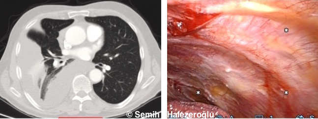
 In a CT scan, areas of pleural thickening or fluid accumulation are detected. The presence of nodular thickening regions is a significant indication of mesothelioma. The image on the side shows CT and thoracoscopy results during the procedure for a patient with mesothelioma in the right pleura, highlighting the affected pleural areas.
MRI is useful for showing the status of the tumor on the diaphragm and heart. If there is suspicion in CT, MRI should be performed. Otherwise, MRI is not necessary.
Mesothelioma that has extended below the diaphragm to the abdominal area or past the heart lining is considered advanced stage.
PET-CT
If a lesion in the pleura raises suspicion of mesothelioma, a PET-CT scan is performed to assess if the lesion is consuming energy. If no increased energy consumption (uptake) is detected in the pleural lesion, the likelihood of it being mesothelioma is very low. PET-CT is also important for determining whether mesothelioma has spread to other areas of the body. No mesothelioma treatment should begin without a PET-CT scan.
Biopsy in Mesothelioma
If fluid is present in the chest cavity, it is collected with a needle (thoracentesis) and the cells are examined. If cells suspicious for mesothelioma are found, a biopsy of the tumor is taken using either an open method or a closed method called thoracoscopy. The pathology exam not only confirms the diagnosis of mesothelioma but also identifies the type. The type of mesothelioma (epithelioid, sarcomatoid, or mixed type) is crucial for determining the appropriate treatment.
Mesothelioma Treatment Methods
Mesothelioma treatment differs based on the stage of the disease and the type of mesothelioma. In many cases, the disease is diagnosed in advanced stages, and chemotherapy is the primary treatment. Pemetrexed (Alimta) has shown significant success. When used in combination with Cisplatin, successful results have been observed. Since chemotherapy may have side effects, a detailed assessment by an oncology specialist is performed before starting treatment.
Heated chemotherapy in mesothelioma, a chemotherapy regimen where drugs are delivered into the chest cavity, has been in use for around 15 years. Long-term results have shown no significant success.
Immunotherapy has been introduced in recent years. Early results are promising, but the role of immunotherapy in mesothelioma needs to be confirmed through extensive scientific research.
In mesothelioma, surgery is only used in certain types of mesothelioma (epithelioid type) and early stages (Stage I and sometimes Stage II).
Radiotherapy (radiation therapy) is often used for organs affected by metastasis, such as the bones and brain. In some cases, radiotherapy can also be applied to mesothelioma itself. However, the application of radiotherapy to a large area should be done carefully, as it may cause adverse effects, and the patient must be monitored closely. Methods such as Cyberknife or IMRT are successful when applied locally to areas where the disease has recurred, as they help reduce the patient’s symptoms.
When is Surgery Applied in Mesothelioma?
Surgery for mesothelioma is only beneficial when performed on a carefully selected group of patients. This patient selection must be done with great care.
- In patients with Stage I and some Stage II disease
- There are scientific studies showing that surgery in cases of mesothelioma with the “epithelioid type” extends the patient’s survival. Therefore, surgical treatment is recommended for this selected group of patients.
- In sarcomatoid or mixed type malignant mesotheliomas or in Stage III and Stage IV patients, surgery does not provide significant benefits!
Mesothelioma Surgery Methods
Surgical treatment consists of two main types:
- Curtative, meaning surgery aimed at completely treating the disease
- Palliative, meaning treatments aimed at improving the patient’s quality of life and making breathing easier during the course of the disease
1. Curative Mesothelioma Surgery
Curative mesothelioma surgeries can be performed in two different ways:
A) Removal of the affected areas of the pleura, diaphragm, and pericardium (Pleurectomy-Decortication). This method is more commonly used today. The image shows a mesothelioma removed through surgery.
B) In addition to the above, the lung is also removed (Extra-pleural Pneumonectomy). This method is applied in rare cases.
The method to be used is decided based on detailed preoperative evaluations and findings during surgery.
2. Palliative Mesothelioma Surgery to Relieve Symptoms
- In mesothelioma patients, if fluid accumulation in the chest cavity causes severe shortness of breath, it must be drained.
- This procedure should be done using the endoscopic (VATS) method in all patients with appropriate general conditions, and a treatment to prevent recurrence of fluid should be applied during the procedure.
- After the VATS procedure, the likelihood of fluid recurrence is very low, but in some patients, fluid may reappear.
Risks of Mesothelioma Surgery
- In curative pleurectomy decortication operations, the risks typically involve prolonged bleeding drainage, air leaks from the lungs, increased heart rate, and heart rhythm disturbances. 3% of patients undergoing surgery may die within 30 days.
- In Extra-pleural Pneumonectomy operations, there is a greater likelihood of requiring a longer stay in the intensive care unit, and 5% of patients may die within 30 days.
- In the palliative VATS pleurodesis procedure, the risks are lower, and the likelihood of death is extremely low.
How Many Days Should I Stay in the Hospital After Surgery?
After curative surgeries, the average hospital stay is 6-7 days, and after VATS pleurodesis, the average hospital stay is 3 days.
Is Radiotherapy / Chemotherapy Needed After Mesothelioma Surgery?
Chemotherapy is generally required. Radiotherapy is not always necessary.
Postoperative treatment decisions are made based on the pathology examination of the surgery and are decided by the hospital’s tumor board.
Mesothelioma is a rare disease, and its treatment should be carried out in centers experienced in this disease.