This article provides information about lung nodules, how they occur, and their symptoms. Based on this information, you can find detailed explanations on the diagnosis of lung nodules, evaluations for benign and malignant nodules, treatments, surgical methods and risks, hospital stay duration after surgery, and follow-up care.
What is a Lung Nodule?
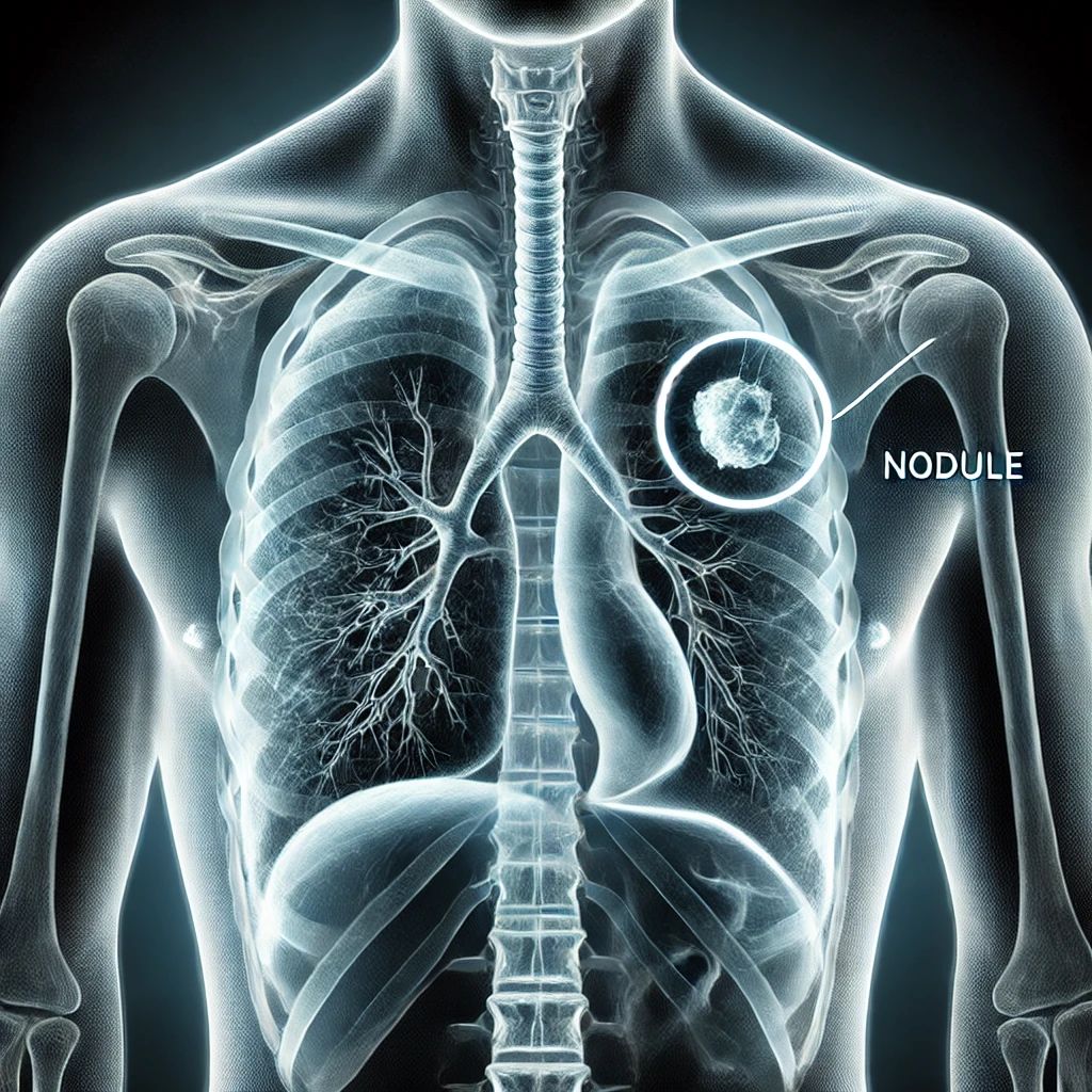
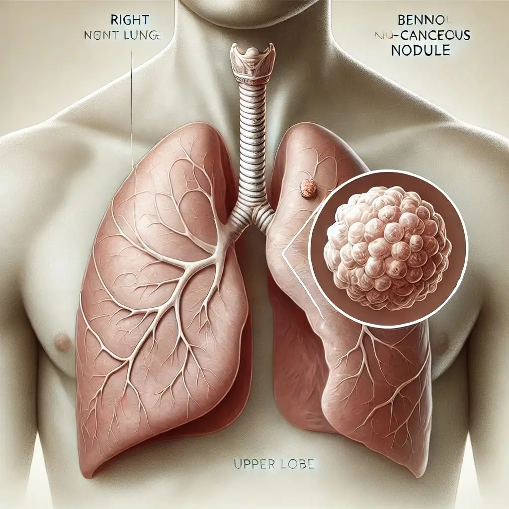
A lung nodule is a lesion in the lung, less than 4 cm in diameter, that may result from residuals of a previous infection, a benign lung tumor, lung cancer, or metastasis of another organ’s cancer to the lungs. Lung nodules are also commonly referred to as “spots on the lungs.”
Often, patients and their families worry excessively about a simple lung nodule. However, experienced doctors specializing in lung nodules can provide the necessary explanations and assessments to alleviate these unnecessary concerns. Therefore, it is important to consult appropriate centers and experienced specialist doctors. Otherwise, unwarranted anxiety can be distressing for everyone involved.
On the other hand, in some cases, a lung nodule with a high probability of being cancerous may not be given the attention it deserves. Neglecting treatment for lung cancer and opting for mere observation can lead to serious consequences.
Frequency of Lung Nodules
Approximately 30% of individuals over the age of 55 who have smoked are found to have lung nodules. Among these individuals, only 1.5% of the nodules turn out to be cancerous. The key is to accurately identify the nodules within this 1.5% cancer risk group.
All our efforts and website resources aim to guide you in understanding the characteristics of benign and malignant nodules.
Radiological Examination of Lung Nodules

Simple chest X-rays are neither sufficient nor appropriate for detecting lung nodules. Lung nodules are identified using computed tomography (CT), and follow-ups, if necessary, are also conducted using CT scans.
Radiological examinations classify lung nodules as follows:
- Solid nodule
- Partially solid nodule or semi-ground-glass nodule
- Ground-glass nodule
- Multiple nodules
- Subpleural nodule
Evaluation of Lung Nodules
Many patients or their relatives send us film reports asking for comments. However, providing a medical opinion based solely on a report is highly inadvisable.
A lung nodule cannot be evaluated merely based on a report or film. Several variables must be considered to make an informed decision:
- Patient’s age
- Smoking history
- Previous illnesses
- Family history of lung cancer
- Whether the nodule was present in prior imaging studies
- Results of needle biopsy, if conducted
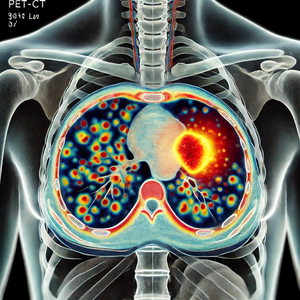
- PET-CT findings, if available
Based on these evaluations, lung nodules are classified as:
- Completely benign nodule
- Low probability of cancer nodule
- High probability of cancer nodule
Benign Lung Nodule
Characteristics indicating that a lung nodule is benign:
- Patient younger than 30 years
- Nodule smaller than 4 mm
- Nodule appearing solid rather than ground-glass on imaging
- Nodule size unchanged in prior imaging studies
- No significant uptake on PET-CT imaging
- No family history of lung cancer
- No prior history of cancer in another organ
- Negative needle biopsy results
- Presence of calcifications within the nodule
- Subpleural nodules are mostly benign.

Benign nodules that are definitively identified require no treatment. In some cases, monitoring is necessary.

To assess whether a lung nodule is malignant, it is insufficient to examine only the nodule itself. The patient’s history and related circumstances must also be considered.
Certain features increase suspicion of malignancy:
- Patient older than 40 years
- Current or past smoker
- First-degree relatives with lung cancer
- History of another cancer
Nodule-specific features raising concern for malignancy:
- Nodule larger than 1 cm
- Ground-glass appearance
- Irregular borders
- Nodule not present in prior imaging
- Increase in nodule size over time
- Increased activity on PET-CT
Which Lung Nodules Should Be Surgically Removed?
Experienced chest physicians or thoracic surgeons may recommend either monitoring or surgical removal of a nodule based on the patient’s history and radiological findings.
Nodules with a high risk of cancer, as described above, should be surgically excised and evaluated by a pathologist to determine the final diagnosis.
Minimally invasive techniques are preferred over open surgery.
Lung Nodule Surgery (Single-Port VATS)
This procedure is performed using VATS, a minimally invasive surgical technique. Under general anesthesia, a 2 cm incision is made in the chest. Through this incision, a 1 cm camera and surgical instruments are used to remove the nodule. The excised nodule is sent for pathological examination during the procedure, and if found to be lung cancer, a lobectomy is performed in the same session. If the nodule is not cancerous, a lobectomy is unnecessary.
Watch the video below to see how this procedure is performed:
After VATS (Minimally Invasive Surgery) for nodule removal, patients are usually discharged within 1–3 days and can resume normal activities three days later. If the nodule is cancerous, requiring a lobectomy, hospital discharge typically occurs in 3–5 days, with work resumption in about 10 days.
The British Thoracic Society published a comprehensive guide in June 2015 for the diagnosis, follow-up, and treatment of lung nodules. Much of the information presented on this page is based on the recommendations from that guide. To access this guide in English, visit https://thorax.bmj.com/content/70/Suppl_2/ii1.full.pdf+html.